

A special chemical process makes all of a mouse’s nerves, tissues, and organs transparent. Scientists in Germany have developed a scanning method that can detect previously unnoticed cancerous tumors, improving the testing of new cancer drugs.
The key element of this new method became transparent mice, albeit lifeless. The method of making a dead mouse transparent was discovered in 2018 by Professor Ali Erturk of the Helmholtz Research Center in Munich, which unites 18 scientific institutions. This is done by chemically removing all fat and pigments from the carcass, making it resemble a flexible plastic toy. The internal organs, blood vessels, and nerve fibers remain intact, but become almost completely transparent. This allows scientists to study the processes taking place in different organs by “staining” them with special antibodies.
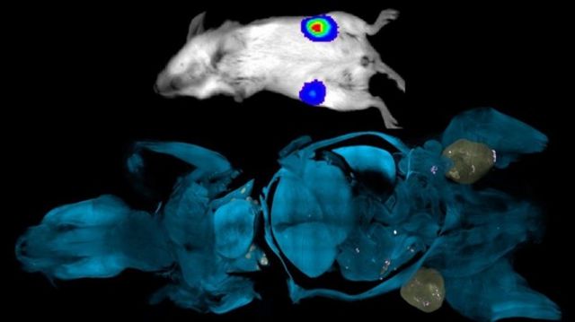
In the lower (blue) image, even tiny cancerous tumors can be seen as white and pink dots. In the normal upper (white) image, only large tumors are visible. It’s no secret that new drugs are often tested on mice. But in this case, scientists claim their method could revolutionize medical research. In the first experiments, researchers were able to detect cancerous tumors in the early stages of formation. According to Professor Erturk, this is extremely important because before clinical trials can be conducted in humans, drugs must prove their efficacy in mice.
“Magnetic resonance imaging (MRI) and positron emission tomography (PET) scans only show large neoplasms,” he explained. “Our method detects tumors at the cellular level.” “The existing drugs can prolong the patient’s life for several years, but then the cancer returns. And this is due to the growth of these very small tumors that went unnoticed,” he adds. Typically, in the laboratory, mice are injected with cancer cells and then scanned periodically to monitor tumor growth. The mice are then given an experimental cancer drug and scanned again to see how it works. The scanning method developed by Professor Ertyurk’s team can only be applied to dead mice, but it provides a clear picture of whether the tumor has progressed or the drug has worked. To do this, mice are made transparent, and only a few mice are needed to determine the effectiveness of the cancer drug being tested.
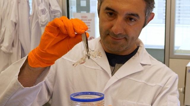
Professor Al Ertürk with a transparent mouse. According to Cancer Research UK’s Rupal Mistry, the proposed unique scanning method has enormous potential to improve understanding of how our bodies work and which specific processes are disrupted during the development of cancer. And although this type of study can only be done on the bodies of mice that have already died, the method can tell scientists a lot about the early stages of disease development. Research in mice is often the starting point for studying processes in the human body. In theory, however, the new method can be used to study tissues from any animal.

Subscribe to our email newsletter and every evening, Monday through Friday, you will receive the most important news of the day, as well as context to help you understand what is happening.